Reproduction
Intro to Reproduction: Physiology and Hormones of the Male and Female
This article covers general information regarding male and female reproductive anatomy.
Quick Scroll
Male Reproduction Introduction
Primary, Secondary, and Accessory Sex Organs of the Male
Female Reproduction Introduction
Reproduction Introduction
Reproduction is the method in which new animals are made and is the most important factor of an animal operation. The unit used to measure reproductive efficiency in an animal is its number of offspring that are born alive. The expectation for this unit is different depending on the species and breed of animal.
For example, a cow that produces one calf per year is considered efficient, while a pig that produces one piglet a year is considered (wildly) inefficient.
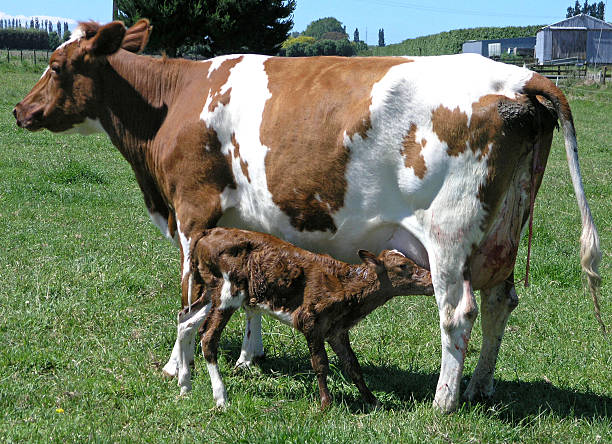
An Ayrshire cow with her calf.
Ensuring reproductive efficiency is incredibly important when it comes to income. The more viable offspring an animal produces, the more income is generated. The offspring of an animal reduces the overhead cost of maintaining said animal, thus generating more profit for the producer.
In summary,
Reproductive Efficiency = Number of Offspring Born Alive
Reproduction = Income
More Offspring = Less Overhead Costs
In order to achieve max efficiency, there are two aspects of reproductive efficiency that a producer must excel in: Genetics and Environment (management).
Genetics is a large determinant of the productivity and health of an animal. The number of offspring produced by a group of animals is directly related to the rate of genetic improvement of said group. The quicker you can produce offspring, the quicker you can make changes across generations through selective breeding. In addition, the more offspring you produce, the more selective you can be about which offspring you will retain to meet your breeding goals.
For example, if you only produce just enough calves to replace cows in a dairy operation, your standards for genetic quality must be lower to retain the quota of calves needed. If you had an excess of calves, you would be able to pick and choose the “best” calves to retain–selling the genetically deficient ones–inorder to improve your operation's productivity. Producing more replacement animals gives you a greater opportunity to select for good genetics. However, it is not recommended to focus solely on quantity. As with all things, you must balance quality and quantity.
Genetics determines the possible productivity of an animal, but its environment is what determines if it can actually reach that potential. The environment of an animal is composed of many aspects: nutrition, cleanliness, housing, seasonality, temperature, other animals, health risks, elevation, stress, etc.
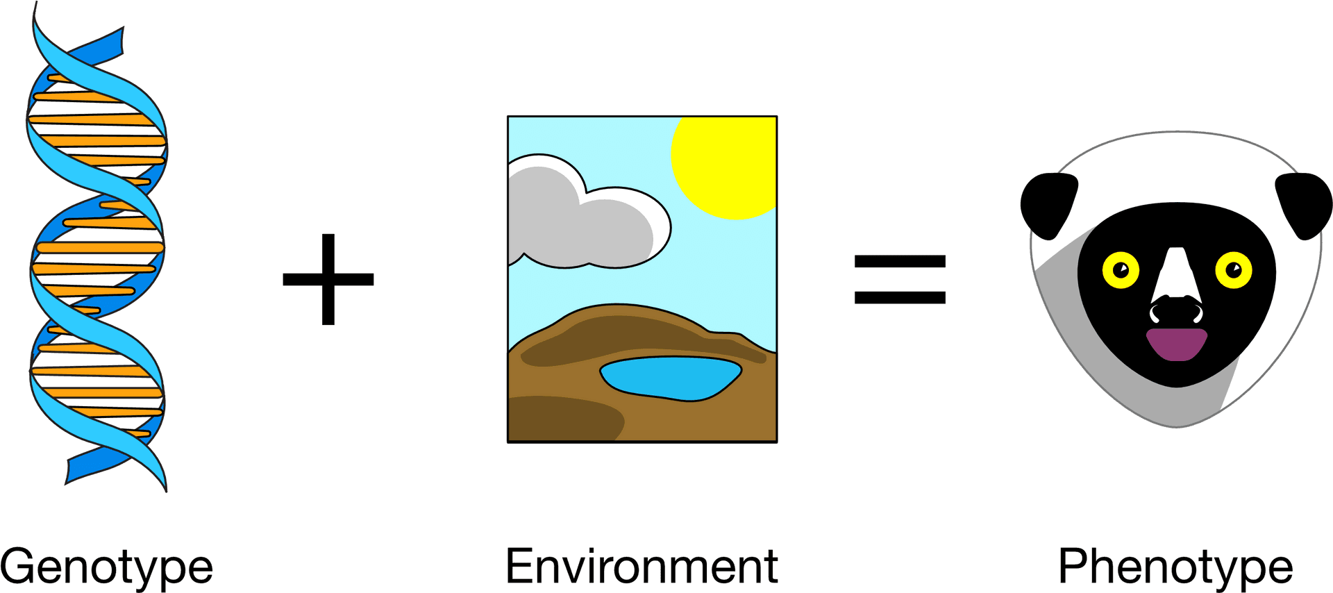
The genetics of animal plus its environment determines its phenotype. Phenotype refers to the way that an animal's genetics are physcially expressed.
When it comes to nutrition, reproductive efficiency is at its best at a body condition score of 5-6. A score of 7-8 is also efficient but is not any different than 5-6 aside from being more expensive to maintain. A score under 5 or above 8 is not efficient, as it indicates malnourishment or obesity, both of which negatively affect reproduction. Ultimately, a female animal will not conceive if they do not have a certain amount of fat. Males produce the most sperm at a moderate weight.
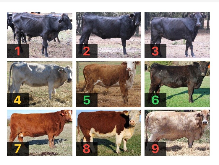
A chart referring to the body condition score of cattle. The lower the score, the thinner the cow, and vice-versa. The scores in green are considered ideal. Image obtained from this article.
Seasonality refers to the time of the year. Animals sense seasonality based on the length of daylight. Some species–such as sheep and horses–are seasonal breeders, meaning that they are only inclined to breed at a certain time of the year. Sheep are short-day breeders, breeding when the days are shorter. Horses are long-day breeders. Because of the difference in gestation length, both species reach parturition (birth) in the spring. Some species, like cattle, can breed year-round. However, conception rates can vary according to season with spring being more favorable than fall. Species that are prone to seasonality can be influenced to breed with modern technology. For example, blue light masks for horses are used to reduce the release of melatonin, simulating a longer light cycle that induces ovulation.

A horse wearing an Equilume mask. The blue light supresses the production of melatonin, tricking the horses brain into thinking the days are longer.
With seasonality also comes changes in temperature. Although seasonality is determined using light rather than temperature, heat and cold still play a part in reproductive efficiency. In a similar manner to its effect on growth, heat negatively affects reproductive efficiency. The cooler it is, the more efficiently an animal can reproduce.
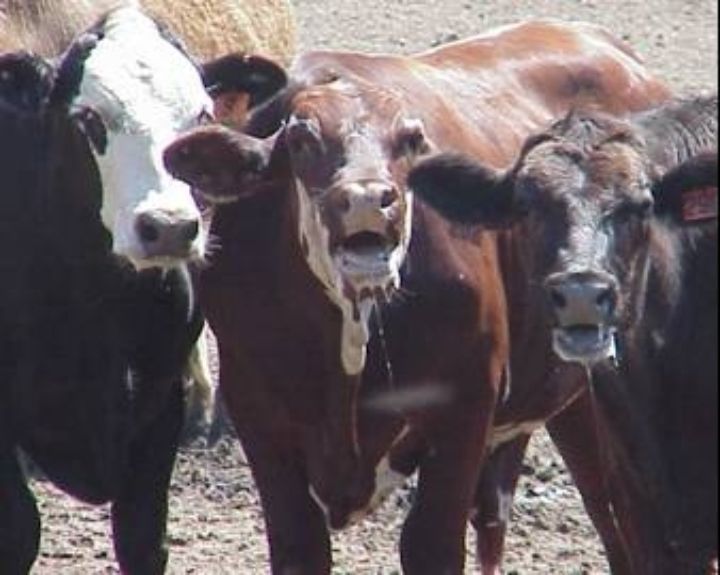
Panting cattle exhibiting heat stress.
Naturally, the ins-and-outs of reproduction varies depending on sex. Below, we will cover both male and female reproduction.
Male Reproduction Introduction
The male has less involvement in the reproductive cycle–thus, it is also less complex to understand male reproduction.
Males are mainly influenced by the hormone testosterone. As a fetus develops, the presence of testosterone influences its sex. If more testosterone is present, male organs are developed, and vice-versa for females. In the case of twins in cattle, a “freemartin” female may develop if the twins are of opposite sex. The influence of testosterone in the male fetus causes malformation of the female fetus' reproductive organs, producing a sterile female.
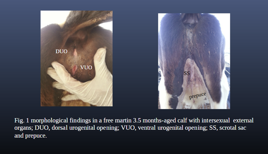
Deformation of a freemartin's reproductive tract.
Although the presence of these hormones in the prenatal stage affects the development of the fetus, it is not until puberty that many characteristics of its sex is developed. In males (and females to a lesser extent), testosterone results in the development of secondary sex characteristics. These characteristics vary depending on species and function (fight or flash). Males are often “showier” than females, displaying colors or behaviors to attract a mate. Think about a peacock versus a peahen. The peacock is more colorful, and “dances” to attract a mate. The color and behavior of that peacock was developed because of the presence of testosterone.
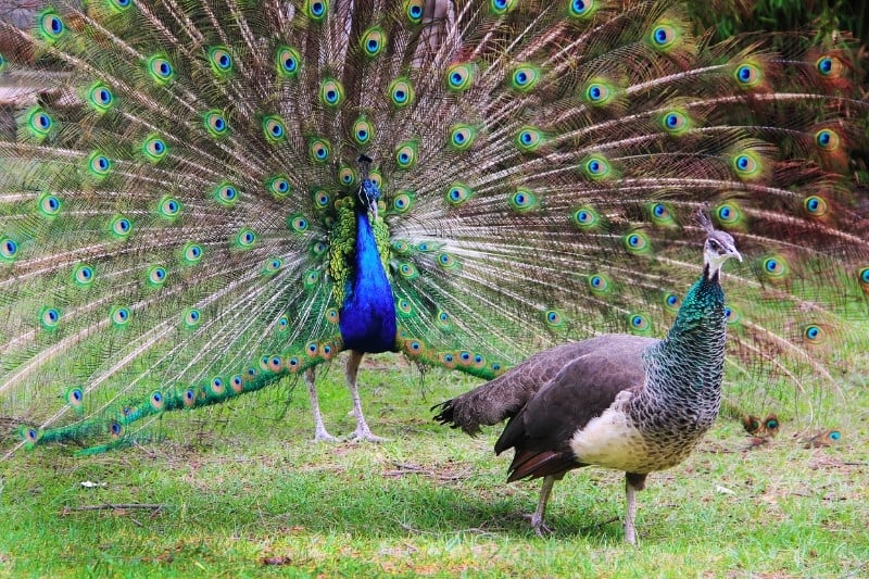
A peacock courting a peahen.
In contrast to flash, fight may also influence male secondary sex characteristics. In cattle, males develop larger horns, thicker necks, and more muscle. These features allow them to compete physically with other males for the right to breed. In livestock breeding, it is preferred for animals to appear “masculine” or “feminine” in accordance with their sex.
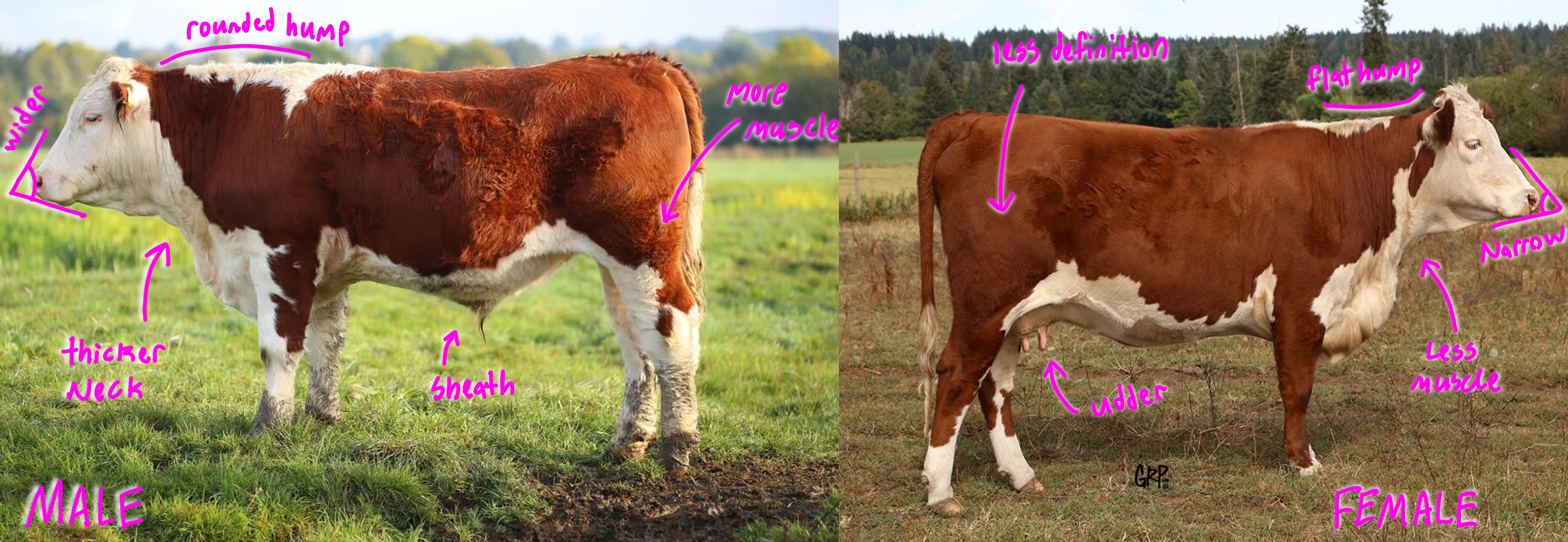
Exhibition of secondary sex characteristics in male (left) and female (right) hereford cattle.
However, not all males are used for breeding. Oftentimes livestock will be castrated to improve the ease of handling and the quality of product. Castration should occur before a male's secondary sex characteristics develop – typically before puberty. Castration is done for various purposes. In males, the testes are where the majority of testosterone is produced. The presence of testosterone typically causes an increase in muscle development. This results in a lack of fat in meat, which reduces palatability. Castrated males tend to fatten faster, deposit more marbling, and grade higher. By reducing testosterone, the behavior of a male is also changed. Typically, males become more docile, making them easier to handle. Naturally, castration also renders a male sterile. This can reduce the need to manage and separate males in a herd. However, the removal of excess testosterone also removes the male's tendency to grow faster.
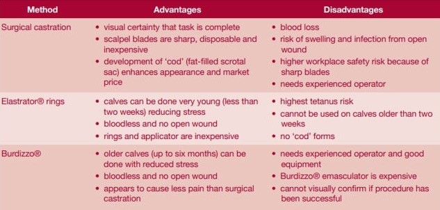
The pros and cons of three main methods of castration: Surgical, Elastrator, and Burdizzo. Surgical castration involves the surgical removal of the testes. Elastrators utilize a rubber band to cut off blood flow to the testes, resulting in necrosis (death of tissue). Burdizzos utilize force to cut off the testes from the body's blood supply, also resulting in necrosis.
Primary, Secondary, and Accessory Sex Organs of the Male
The male's reproductive system is comprised of three categories: Primary, secondary, and accessory sex organs.
The primary sex organ of the male is the testicle (testes for plural). The testicle of an animal can be internal or external. For example, a chicken's testes are internal, while a bull's are external. The difference between external and internal – aside from position – is the temperature at which the animal's sperm cells can thrive at. For internally-positioned animals, the sperm must be kept at a higher temperature. For externally-positioned animals, the sperm will die if kept at body temperature for too long, and thrive at slightly cooler temps. In order to regulate their temperature, the testes may extend and retract from the body. Upon extending, they become cooler—and vice-versa.
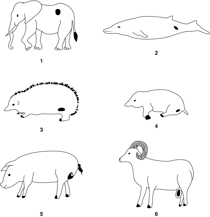
Testicular placement in various animals. Animals one through five are internal. Animal six is external.
The size of an animal's testes affects it's reproductive performance. The optimal size is dependent on the intended use of the animal. For terminal males (males headed to slaughter), the size doesn't matter. For non-terminal males (males intended to be used for breeding), a larger size is more desirable. This is not really due to the increased production of sperm and testosterone in the male – rather, the size of the testes also helps to determine the size of their female offspring's reproductive tract. Because a larger tract is more efficient in females, larger testes are optimal for their sires. The size of a male's testicles also affects the speed at which the male hits puberty. The larger the testes, the sooner puberty is reached.
So, what exactly is the function of the testes? This organ produces the male gametes. Gamete is the term used to refer to sex cells. The sex cell, or gamete, of the male is the sperm. As the male's primary sex organ, the testicle is the site of spermatogenesis in males.
Spermato = sperm
Genesis = to produce
Thus,
Spermatogenesis = the production of sperm
Spermatogenesis occurs in the seminiferous tubules of the testicle. Unlike females—who are born with all of the eggs they will ever produce—males continuously regenerate their sperm cells after they reach puberty. The testicle has endocrinal (hormonal) function, producing the male hormone testosterone in its interstitial cells.
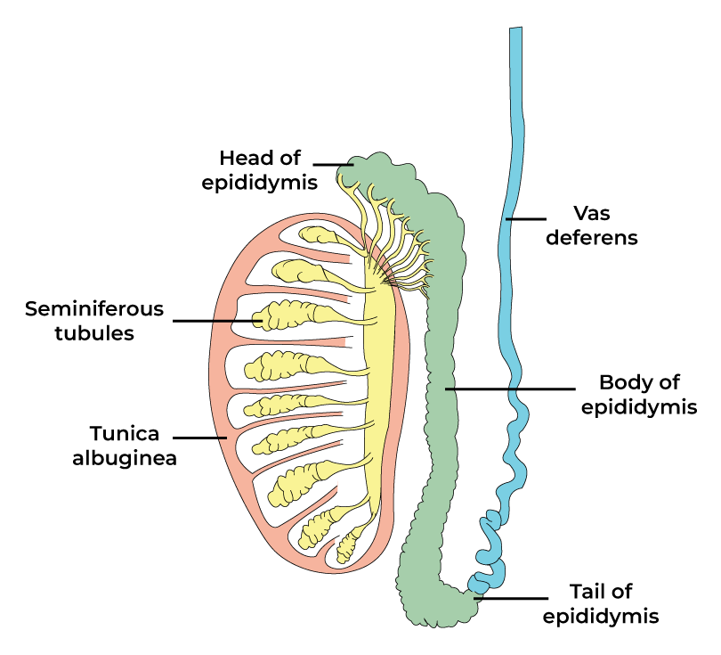
Diagram of the testicle. Sperm is formed in the yellow lobed structures.
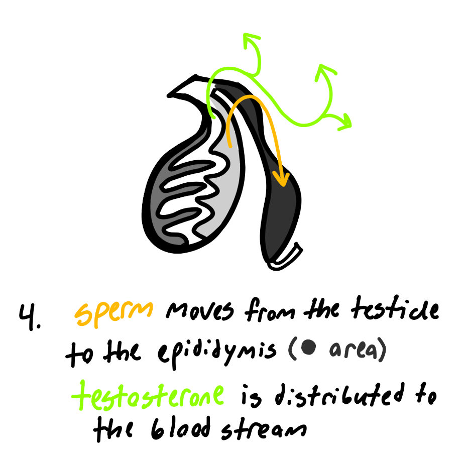
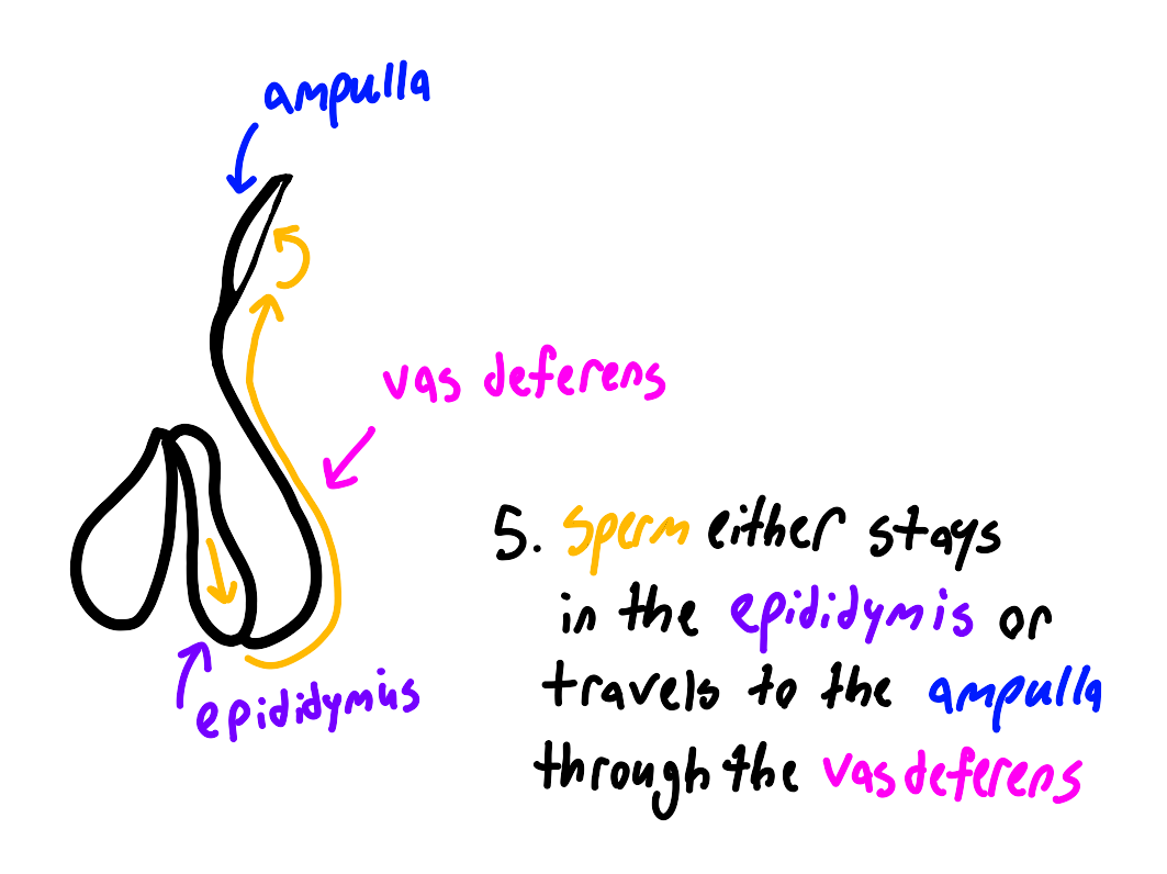
The hormone testosterone is then dispersed into the blood stream, where it is distributed into the body. As for the male's sperm, it is carried from the tubules of the testes into the epididymis, starting the involvement of the male's secondary sex organs. The epididymis acts as a storage area for sperm to be held while it matures.
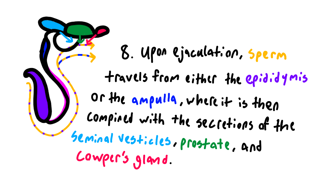
Upon ejaculation, the sperm is moved from the epididymis through the vas deferens. The vas deferens acts as the sperm's “pathway” outside of the body, ending at the urethra. However, this pathway may be interrupted by the ampulla. The ampulla is not present in all species. Species may either be considered fast or slow ejaculators. In fast ejaculators (bull, stallion, ram, and buck), the ampulla acts as a temporary storage area for sperm—sort of like a “shortcut,” reducing the time needed for the sperm to exit the body. In slow ejaculating species (boar, dog, and human), the ampulla is not present, and the sperm travels directly from the epididymis to the vas deferens.
In the case of humans, one may undergo a vasectomy. In a vas-ectomy, the vas deferens is severed. This prevents the sperm from reaching the urethra, thus preventing ejaculation. However, this method of sterilization is not done in pets or livestock. Humans are vasectomized rather than castrated in order to retain the testosterone production in the testes. If the testes were to be removed, a male would suffer the effects of a lack of testosterone. Although vasectomizing an animal is entirely possible, it's actually not the preferred method. It requires more surgical precision, making it more expensive and labor intensive. In addition, the removal of testosterone production is one of the goals of castration. Without removing the testes, livestock and pets would still produce testosterone, which is undesirable (due to the physical and behavioral effects mentioned earlier–as well as the possible health risks in pets) unless your goal is to breed the animal.
Now that we have covered the testes, we can discuss what covers the testes. The scrotum is what contains and protects the male's testicles. It aids in temperature regulation through the cremaster muscle, retracting and extending the testes to maintain their temperature at 4 to 5 degrees Fahrenheit cooler than the body's temperature.
Males may have deformations concerning the scrotum and testes that occur upon the “dropping” of the testes during puberty. In the case of a cryptorchid, both testes are retained inside the body cavity. This produces a sterile male, as the sperm dies due to the high temperature. In the case of a monorchid (mono = one), only one testicle drops, and the other is retained inside the body. Monorchids are fertile – but it is not recommended to breed them, as their offspring may have reproductive issues.
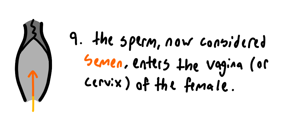
The male also has various accessory sex glands. One of these is the prostate. The prostate nourishes and stimulates the activity of sperm through secretions. Sperm, upon receiving these secretions in addition to those of other glands, becomes semen. To reiterate, sperm refers to the cells themselves, while semen refers to the mixture of sperm and accessory secretions. The fluid contained in these secretions helps the sperm to swim. There is also an enzymatic secretion from the vesicular glands (seminal vesicles) that neutralizes any urine residue that may harm sperm. The bulbourethral gland also serves to neutralize urine through enzymes, as well as adding volume to the ejaculate. In boars, a thick gel is formed by this gland that plugs the cervix in the sow. All of the above accessories open into the urethra.
Prostate = excretes fluid that helps mobilize and nourish sperm
Vesicular glands = produces enzymatic fluid to neutralize urine
Bulbourethral gland = produces enzymatic fluid to neutralize urine. Also adds volume to sperm, especially in the case of boars.
The urethra is a muscular canal along the length of the penis that is used as a pathway from the bladder and vas deferens. It is used to excrete urine and ejaculate.
The male's prostate sits near the urethra and may cause urination issues if it becomes inflamed. Usually this occurs in senescence (old age). These issues are not commonly seen in livestock as they are typically slaughtered before then.
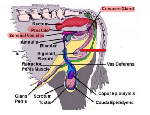
Diagram showing the location of the accessory organs and glands (highlighted in red) of a bull.
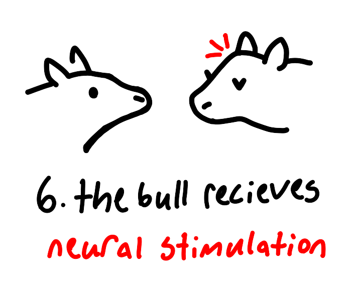
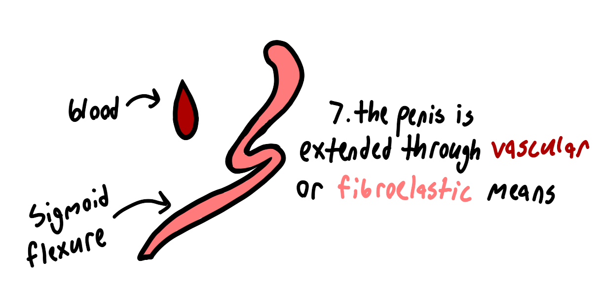
Another part of the male's reproductive tract is the sigmoid flexure. This muscle is an internal “S” shaped structure. It's function is to maintain erections through contracting and relaxing. Upon relaxing, the penis is extended, and vice-versa. This muscle is affected by neural stimulation. If a male has a sigmoid flexure, they have what is considered a fibroelastic penis. This includes the bull, boar, and ram.
Species that do not have this muscle are known as vascular species. Vascular is a term used to refer to blood. If a male has a vascular penis, it means that their erection is maintained through the flow of blood. The flow of blood to the penis is also affected by neural stimulation. Vascular species include the stallion, dog, and man.
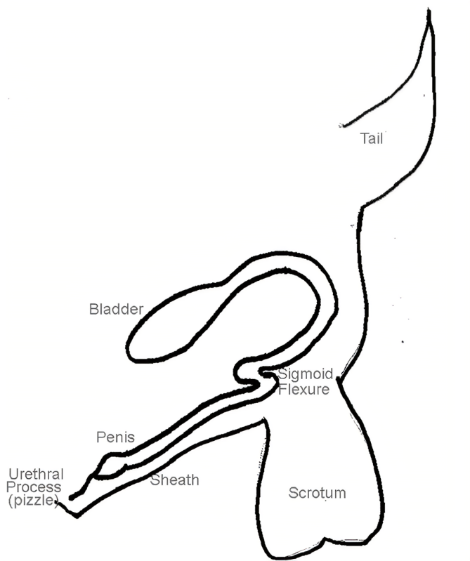
Diagram showing the location of the sigmoid flexure.
Aside from the method of an erection, males of different species can also have differently shaped penises. The penis is considered the male's copulatory organ, or the organ that allows for sexual intercourse. In livestock, the most notable variations in shape are that of the boar and the ram.
Boars have what is considered a “corkscrew” apparatus. This is used to enter the longer cervix of the sow. Because of this adaptation, boars ejaculate a lower volume of semen than other species, as the semen does not need to travel as far.
Rams exhibit an extension of the urethra, extending beyond the tip of the penis. This extension is sometimes cut off in order to reduce complications with urinary calculi. Despite this, the ram remains fertile.
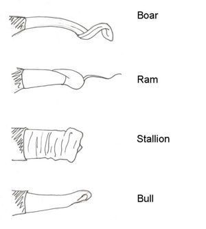
Diagram showing various shapes of livestock penises.
Male Hormones
All of the systems above are effected by the presence of various hormones. These hormones are produced and are then dispersed around the body. Upon which, they are picked up by receptors in cells where they then facilitate various processes. In summary, hormones are chemical messengers that regulate bodily functions. In a similar manner to female animals, the male's reproductive system is effected by three main hormones: GnRH, LH, and FSH.
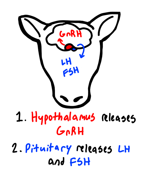
GnRH (gonadotropic releasing hormone) is produced in the hypothalamus. It stimulates the release of LH and FSH from the pituitary gland.
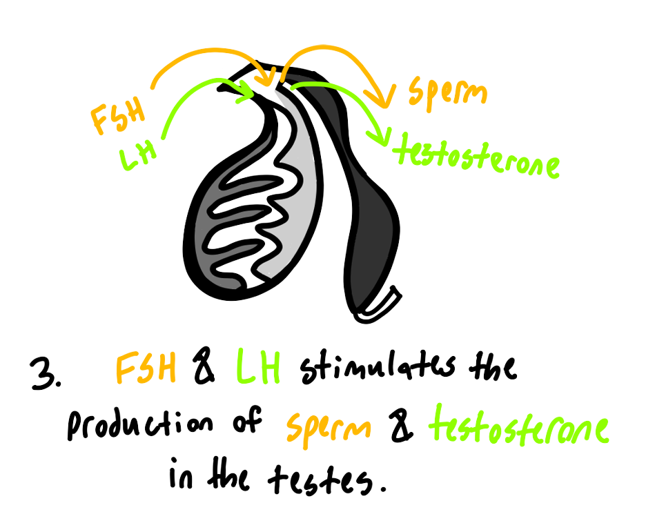
Speaking of, LH and FSH are produced in the pituitary gland. FSH (follicle stimulating hormone) binds to the seminiferous tubules of the testicle and stimulates the production of sperm. LH (luteinizing hormone) stimulates the production of testosterone in the testes.
Because the hormones act as the facilitators of reproductive processes, any issues with the amount of hormones produced can cause a myriad of health problems.
Male Reproduction Summary
Below is a series of images that chronologically explains the reproductive process of the male.
Female Reproduction Introduction
The female reproductive system is a tad more complicated than the male's. We will begin with a quick overview of the entire system, and then we will focus on its various parts.
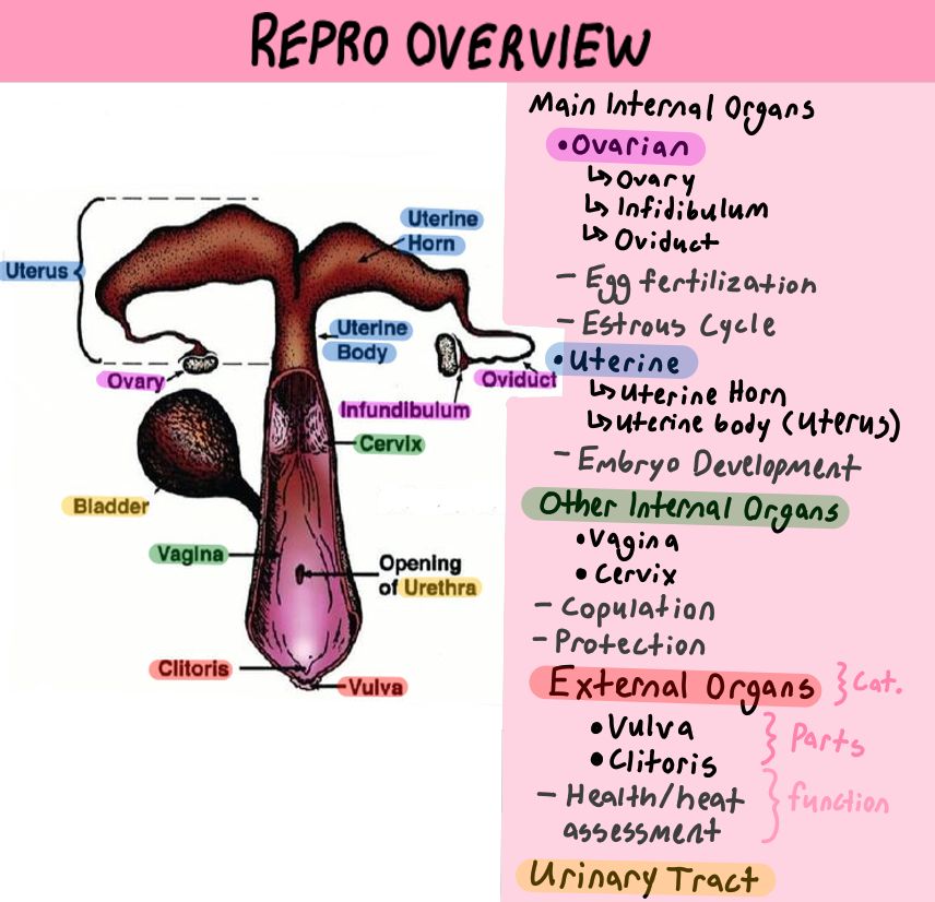
A diagram showing an overview of the female reproductive anatomy. Each part will be covered below.
The Ovary
Just as we did with the male, we will begin with the female's primary sex organ, the ovary. The ovary is the most complicated aspect of the female reproductive system. It produces ova, or eggs, as well as the hormones estrogen and progesterone. Unlike males, a female is born with all of the gametes (eggs) that she will ever produce.
Cell terminology, such as gamete or germ cell, can get kind of confusing. To clarify, a germ cell undergoes meiosis, splitting in half to form four gametes. In males, all four gametes typically form functional sperm. In females, only one or two of the gametes formed becomes an egg. The rest form polar bodies that are quickly degenerated. After they are formed, a gamete is then combined with that of the opposite sex through copulation to produce a fertilized zygote. A zygote then undergoes several stages, eventually developing into a fetus.
Now that we have covered what an ovum (egg) is, we can discuss its journey from the ovary to fertilization.
The ovum begins in the primary follicles of the ovary where it is housed until the body sends hormonal signals to begin the estrous cycle. The primary follicles act in a similar manner to a male's epididymis. It is where the ovum matures until it reaches the Graafian stage, upon which it is ready to be released. During this stage, the ovum is housed in a fluid-filled follicle. This follicle becomes bigger as it matures, and the walls surrounding it become thinner as a result of more fluid being trapped.
Once the follicle is fully into the Graafian stage, it is then just a matter of time before the follicle ruptures. Upon rupturing, the ovum is shot out of the follicle alongside the fluid. This is referred to as ovulation. After ovulation, the ovary is left with a wound-like opening called the corpus luteum. The corpus luteum, meaning “bloody body,” then develops over the next few days. It takes on a yellow color due to the accumulation of lipids needed to produce progesterone.
If the animal conceives, the corpus luteum will continue to produce progesterone, which is needed to maintain the pregnancy. If an animal's CL does not produce enough progesterone, the pregnancy will be aborted. This is commonly an issue seen in horses. This issue may be resolved through progesterone-releasing implants or food additives.
If an animal does not conceive, the corpus luteum will be exposed to prostaglandin, which signals it to regress, ceasing the production of progesterone. Upon regressing, the corpus luteum scars and becomes pale, now being referred to as a corpus albicans, or “white body,” marking the end of the cycle. Upon the next estrous cycle, a new follicle will begin to grow, thus beginning the ovum's cycle over again.
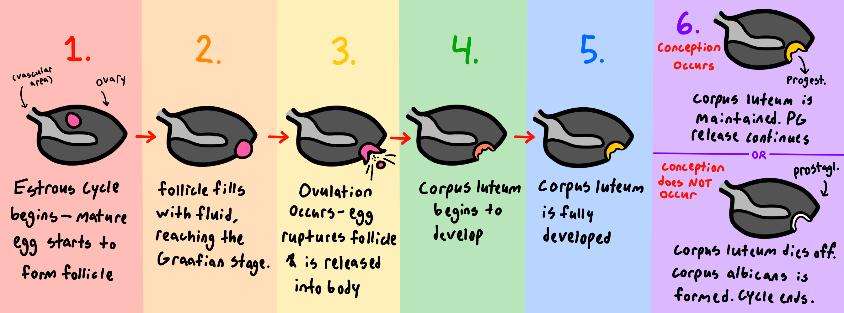
A diagram showing the full cycle of the egg through the ovary.
Anestrous
Before puberty or after parturition, an animal will be in a stage of anestrous. Anestrous means that they will not have an estrous cycle (“an” meaning lack of–remember it as “lack of an-estrous cycle!”) In seasonal breeders such as horses and sheep, anestrous occurs if the proper light requirements are not met. Malnourishment will also cause anestrous. Obese animals may cycle, but they commonly exhibit issues with hormonal imbalances and irregularities, making conception difficult.
While humans will reach menopause at an older age, livestock do not have a point in life where they permanently cease their estrous cycle. Rather, the lack of estrous in livestock reaching senescence is referred to as senile anestrous. Older livestock may have periods of senile anestrous followed by a normal estrous cycle. However, this is not an issue that is commonly seen in livestock, as they are normally slaughtered before reaching senescence.
Fertilization of the Ovum
Now that we've discussed how the egg leaves the ovary, we can move on to how it becomes fertilized.
Once the ovum is released from it's follicle in the ovary, it becomes free-floating. The funnel-like infundibulum works to “catch” the egg, preventing the ovum from becoming lost in the body cavity. The ovum then travels down the infundibulum and into the oviduct (also referred to as the fallopian tubes). There, the egg encounters semen and becomes fertilized. It typically takes millions of sperm cells to fertilize one egg due to the low chances of an encounter.
Once the egg is fertilized, it begins to float down the uterine horn of the animal, eventually becoming attached to the uterine wall. Here, a placenta is formed, and the zygote receives nourishment, eventually developing into a fetus. Litter-bearing animals such as pigs can release many ova at once. Non-litter-bearing animals usually only release one or two eggs at a time. Because of the difference in litter size, the uterine horns of litter-bearing animals are longer. Non-litter-bearing animals have shorter horns. When it comes to artificial insemination, the semen is typically deposited in or just before the uterine horns of the animal. To learn more about reproductive technology, please visit this article!
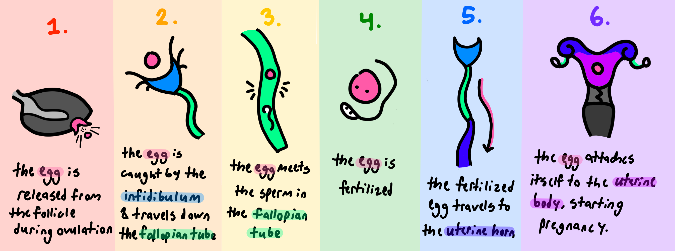
A diagram showing how an egg becomes fertilized.
Other Female Internal Sex Organs
That was a lot, wasn't it? Thankfully, the rest of the female's reproductive tract is a little simpler. Now, we will move on to the other internal sex organs.
We have mentioned the uterus. The uterus is where the fetus stays and develops until parturition. Before the uterus is the cervix. The animal's cervix is a thick section of cartilaginous tissue that serves to protect the uterus. During heat (estrus) the animal's cervix opens, allowing for the passage of sperm for fertilization. Outside of heat, as well as upon the attachment of a zygote to the uterine wall, the cervix closes and becomes impenetrable. This acts as a sort of “barrier” to keep out anything that may harm the animal's reproductive organs or a potential fetus. The cervix only opens on two occasions: the event of estrus and during parturition. During estrus, the cervix opens to allow sperm to pass through. In the process of AI, the cervix is the most difficult barrier to get the rod through because of its firm, mountain-like structure. Sperm must pass through this structure to get to the egg. In the case of parturition, the cervix opens to allow the developed offspring to be born. The opening and closing of the cervix occur because the cervix is incredibly responsive to hormonal stimulation. Later, we will discuss the hormones that affect the cervix.
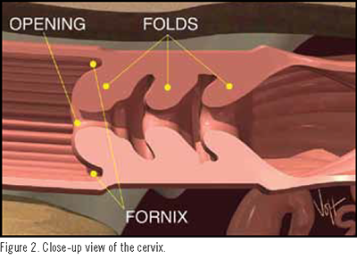
A close-up view of the cervix. To the right of the cervix is the uterine body.
The last internal sex organ is the vagina. The vagina is the copulatory organ of the female, meaning that it is where copulation (sex) takes place. During sex, the male ejaculates into the vagina using his copulatory organ, the penis. The sperm then travels past the cervix, up the uterine horn, and into the oviduct to fertilize the egg.
Female External Sex Organs
Outside of the body is the vulva and clitoris. The vulva refers to the external portion of the vagina. It has little use in the actual process of reproduction but can be a useful indicator of when an animal is in heat. Upon entering heat, the vulva will swell with blood and or change its color. This is especially true in swine. It may also help a producer to assess the health of a female's reproductive organs. If the vulva is small, misshapen, oddly colored, or leaking discharge, it may indicate that an animal is either not fully developed or that it has an issue with the rest of its reproductive tract.
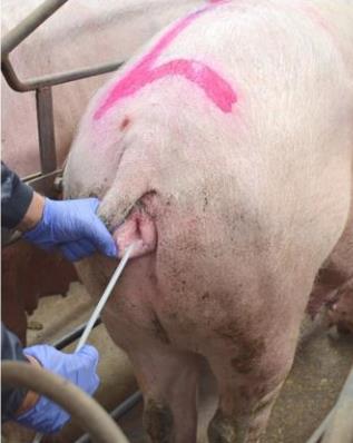
This image shows a pig in heat undergoing AI. Note the swelling of the vulva.
Sitting near the vulva is the last external sex organ known as the clitoris. The purpose of this organ in livestock is unknown. Studies conducted have shown that stimulating the clitoris has no effect on conception rates, making its function in livestock obsolete.
Female Hormones
Now that we understand the physical aspects of the female reproductive tract, we can dive into how hormones affect said features. Like the male, the female's hormone production begins in the hypothalamus. The hypothalamus produces gonadotropic releasing hormone (GnRH). This hormone then stimulates the anterior pituitary to release luteinizing hormone (LH) and follicle stimulating hormone (FSH). Follicle stimulating hormone (FSH) does as it says, it induces the development of follicles on the ovary. The more FSH an animal produces, the more follicles will develop. This can be seen in processes like superovulation, where a female is given excess FSH, resulting in her developing more follicles than she normally would. Read more about superovulation and embryo transfer in my reproductive technology article!
Luteinizing hormone (LH) helps ovulation to occur. LH induces Graafian follicles to rupture and the corpus luteum to develop.
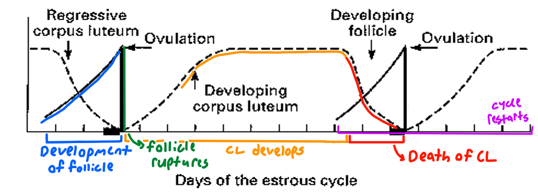
This chart demonstrates the events of the estrous cycle.

This chart demonstrates the fluctuation of hormones as they relate to the events of the estrous cycle.
The last hormone produced in the anterior pituitary is prolactin. Prolactin (pro-lactin, with lactin referring to milk), stimulates lactation, or the production of milk. This hormone also discourages an animal from re-breeding during lactation. However, some animals such as horses can still breed while lactating.
The words anterior and posterior are directional terms used to refer to relative position. Anterior means in front. Posterior means behind. For example, a cow's head is anterior, while its tail is posterior. I remember it as anterior referring to antennas, and posterior being a fancy term for the buttocks.
Oxytocin, produced in the hypothalamus and stored and released by the posterior pituitary, causes the milk to be pushed down from the udder into the animal's ducts, allowing it to be accessed by the offspring through suckling. This is called milk letdown. Oxytocin also stimulates uterine contractions. These contractions facilitate the birth process, pushing material outside of the uterus. It is especially important in ridding of the leftovers of the fetus, such as the placenta. Horses are particularly at risk if this material is not removed within hours of parturition. In this case, they may be given extra oxytocin to help induce more contractions. In poultry, oxytocin induces egg-laying.
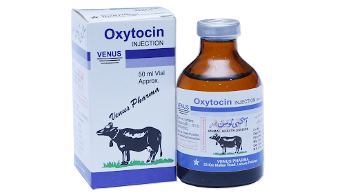
An oxytocin injection.
The gonadal (ovarian) hormones are hormones that are produced by the ovaries. These hormones help facilitate the estrous cycle as well as parturition.
Estrogen is produced from follicles in the ovary. It is known as the primary female hormone and is responsible for the development of both physical and behavioral secondary sex characteristics. Estrogen also causes duct growth in mammary glands, induces estrus (heat), prepares the uterus for pregnancy, and increases the motility of the uterus and oviducts during estrus.
Progesterone is produced from corpus lutea. Progesterone prepares the uterus for pregnancy—preventing ovulation, encouraging uterine growth, and stimulating the development of milk producing tissue.
The last gonadal hormone is relaxin. Relaxin is produced by the ovaries or placenta, and, as the name suggests, causes relaxation of cartilaginous tissue and ligaments in the pelvis. This helps to ease parturition. Sometimes, heifers that experience dystocia will be given excess relaxin to increase their flexibility.
Parturition
All of the factors above work together towards one goal: successful parturition of a live offspring. As mentioned above, parturition is the term used for birth. If a female experiences issues with parturition, it is referred to as dystocia. Dystocia can occur for various reasons, one of which being abnormal fetus presentation. Presentation refers to the position of the fetus (legs-first, head-first, etc.). The smaller and more streamlined an offspring is, the easier parturition becomes. Below are some charts showing various calf positions. The ones labeled as abnormal are undesirable and may require human assistance in repositioning the calf by hand or a cesarean section (c-section) by a veterinarian to resolve.
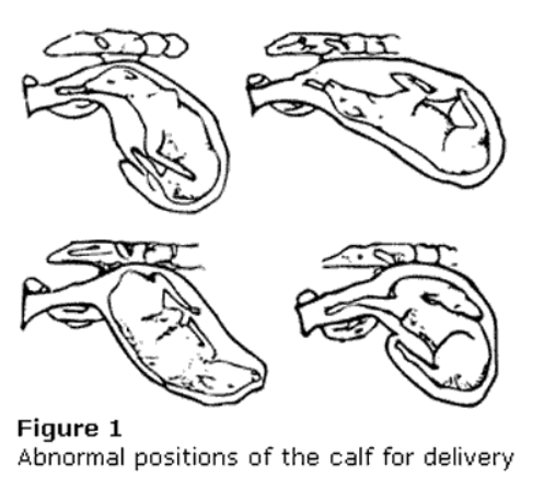
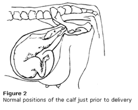
Two diagrams showing abnormal (left) and normal (right) calving positions.
With a fetus also comes a placenta. The placenta acts to protect, nourish, and transfer hormones to and from the fetus during its development. It's kind of like the fetus' communication hub with the mother. Nutrients from the mother pass into the placenta, and waste from the fetus passes to the mother. The placenta protects the fetus from shock and from the transmission of bacteria.
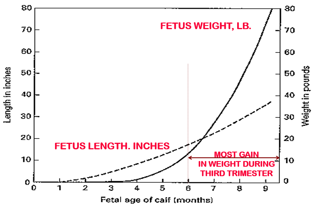
This chart shows the rate of gain in weight (solid line) and height (dashed line) of a cattle fetus during pregnancy. The fetus gains the most weight during the third trimester, or last third, of the pregnancy.
There are two types of placentae, diffuse (mare and sow) and cotyledonary (ewe and cow). The larger the placenta, or the more cotyledons that are present, the more potential the fetus has. As mentioned previously, it is essential that the placenta is also passed during parturition. In the case of cattle, the placenta may be passed within 12 hours of birth. For horses, it must be passed within a few hours.

The difference between a diffuse (left) and cotyledonary (right) placenta.
And thus marks the end of this article on the basic reproductive physiology of the male and female. Congrats on making it through!